Clinical focus: cool the burn, warm the patient - a refresh on treating burns

Fire photo OPT

Globally, about 11 million people seek medical treatment, and 300,000 die, from burns each year.

And since January, our service has received more than 1,500 emergency 999 calls to people suffering from burns or explosion related injuries.

But how should we deal with burns?
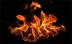
About burns

  • A burn wound has three zones: a zone of coagulation that contains the destroyed cells, a zone of stasis which contains injured cells that may then die, and a zone of hyperaemia which has minimal thermal damage.
  • Pain from a burn injury comes from nerve endings in burnt skin being exposed; this is why covering a burn in cling film or similar can provide quick pain relief and so should be used, but adequate analgesia is also really important. With burns, it’s also vital to take whatever steps you can right from the outset to minimise the risk of infection.
  • Fluid loss from the surface of the burns happens because of vasodilation and changes in the blood vessel walls. The fluid loss is proportional to the extent of the burn and will be present for around 36 hours after the injury.
  • Burn shock is slower in onset (more than one hour) than hypovolemic shock which may be associated with other injuries.
  • Hypothermia, especially in children, can occur with over-zealous cooling. Remember - cool the burn, warm the patient.  
  • Early burn related deaths are often associated with airway obstruction and severe inhalation injury. Later deaths are often precipitated by sepsis with a sustained inflammatory response, acute lung injury, renal impairment, and myocardial depression leading to multiple organ failure.
  • A poorer prognosis is associated with burns to the face (often linked with smoke inhalation), hands or feet, eyes, ears and perineum. Burns of these areas, regardless of size, may need managing in a specialist burns unit.


Types of burns

Scalds: scald burns are very common in children – 85% of children that are admitted with burns have scald injuries and are under three years old. Most often caused by hot liquids, they can also frequently be seen in the elderly or those using drugs and alcohol, and are sometimes associated medical crises like hypoglycaemia or CVA.

Flame burns: these are seen mainly in patients from house fires, especially if their clothes have caught alight. Accelerants used to start bonfires or BBQs often lead to flame burns and may also be associated with airway burns.

Chemical burns: chemical burns commonly follow domestic and industrial accidents, but occasionally happen as a result of as self-harm or deliberate attacks. Specialist advice will need to be gained to deal with some situations.

Electrical burns: these may follow a low or high current source; low current characteristically produces a small deep burn at the entry and exit points with variable tissue damage between them. Depending on the anatomical path of conduction, there may be cardiac dysrhythmias.

Contact burns: result from skin contact with a hot object and depend on the nature and temperature of the heat source, and the length of contact.


Burn depths

The anatomical depth of a burn can be divided into:

  • simple erythema – a superficial burn with no skin loss. Skin is red and tender and heals within five to 10 days with no scarring.
  • superficial partial thickness burns – painful, with superficial blistering healing in 10-14 days.
  • deep partial thickness burns – thick walled blisters with granular white skin and pinpoint red mottled areas. Associated pain may be limited because of damage to the skin adnexae.
  • deep full thickness burns – producing white leathery charred skin with no sensation. These burns commonly follow prolonged contact with a burning agent or dry heat. 

Surface area may be assessed using the rule of nines in adults, adapted for children. The palm of the hand in adults (with the adducted fingers included) equates to approximately 1% body surface area. Do not include areas of erythema.


Factors contributing to poor outcomes

  • Inhalation injuries can be a major cause of death due to toxic substances, like carbon monoxide, causing systemic poisoning.
  • Age is everything; children under five years old are at the highest risk as the surface area of the burn is bigger in relation to their total body size. The elderly are also at a higher risk due to poorer healing potential.
  • Dependant on the circumstances, there can be associated injuries; if the patient has fallen or jumped from height during a building fire for example, or impact injuries in road traffic accidents. Electrical injuries, including arrhythmias, should be considered if you’re dealing with electrocution burns.
  • Underlying significant co-morbidities including ischaemic heart disease, diabetes mellitus, steroid therapy and immunocompromised patients can all lead to poor outcomes in burns patients.


Assessment and management

Scene safety: Ensure the scene is safe for you and the patient. If safe to do so, remove the heat source and stop the burning process and remove hot and/or burnt clothing.

Start to prepare for cooling: Remove any constricting jewellery from the patient, and irrigate the burn with copious amounts of water (minimum of 15 minutes for chemical burns, and a maximum of 20 minutes for all other burns to avoid hypothermia) as soon as practicable. This can still be effective up to three hours after the injury. Alkali burns (some cooking oils) require prolonged irrigation continue to definitive care. Do not use ice or iced water.

Assess C – ABCDE:

Key points for the burns patient –

C - check for catastrophic haemorrhage.

A – airway patency as early intervention may be required with inhalation burns.

B – administer oxygen via a non-rebreathing mask. Sp02 readings may be false due to carboxyhaemoglobin. If patient is wheezing due to smoke inhalation, administer nebulised salbutamol.

C - fluid resuscitation if indicated.

D – assess the size of the burn, check limb perfusion and function distal to burn site.

E – check all areas, remove clothing (but remembering to consider hypothermia). Use small sheets of clingfilm (do not wrap the limb) as required and consider further pain relief options.

  • Consider direct access to burns centre.
  • ATMISTER pre alert.
  • Contact 202 critical care desk.


Documentation

Remember to record:

  • how the patient was burned
  • the time the burn occurred and how long the patient was exposed to source of burning
  • temperature of the source of burning (e.g. boiling water, hot oils etc.)
  • time and volumes of infusions

Further reading – AACE (JRCALC) clinical practice guidelines page 239-247.

Published 2nd November 2014 

0 Comments
Leave a Comment
Name (required)
Email Address (required, never displayed)
Enter a message

(all comments are moderated - your submission will be posted on approval.)